[View 38+] Covid 19 Virus Electron Microscope Image
Download Images Library Photos and Pictures. Electron microscopy (SEM and TEM) images of SARS-CoV-2 - Covid 19 Electron microscopy images reveal how an anti-cancer virus interacts with tumour cells - ecancer T4 bacteriophage via electron microscope | Electron microscope, Microscopic, Microscopic photography Modern Uses of Electron Microscopy for Detection of Viruses | Clinical Microbiology Reviews

. The deadly knife of the influenza virus made visible Correlative Scanning-Transmission Electron Microscopy Reveals that a Chimeric Flavivirus Is Released as Individual Particles in Secretory Vesicles Cryo-electron microscope research reveals structure and mechanism of Bluetongue virus | UCLA
 A) Electron microscope image of virus particles forming a crystalline... | Download Scientific Diagram
A) Electron microscope image of virus particles forming a crystalline... | Download Scientific Diagram
PLOS ONE: Different Regions of the Newcastle Disease Virus Fusion Protein Modulate Pathogenicity
 Pin on Microscopically Magnified
 MERS-CoV Photos | CDC
Focus On: SARS-CoV-2
 COVID-19 infection triggers growth of arm-like tentacles in cells, microscope images show
 Electron microscopy (SEM and TEM) images of SARS-CoV-2 - Covid 19
 A New Phlebovirus Associated with Severe Febrile Illness in Missouri | NEJM
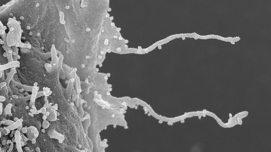
 Lab uses electron microscopy to visualize influenza invasion | Newsroom
 MERS-CoV Photos | CDC
 Health: Coronavirus Prevention Tips | Voice of America - English
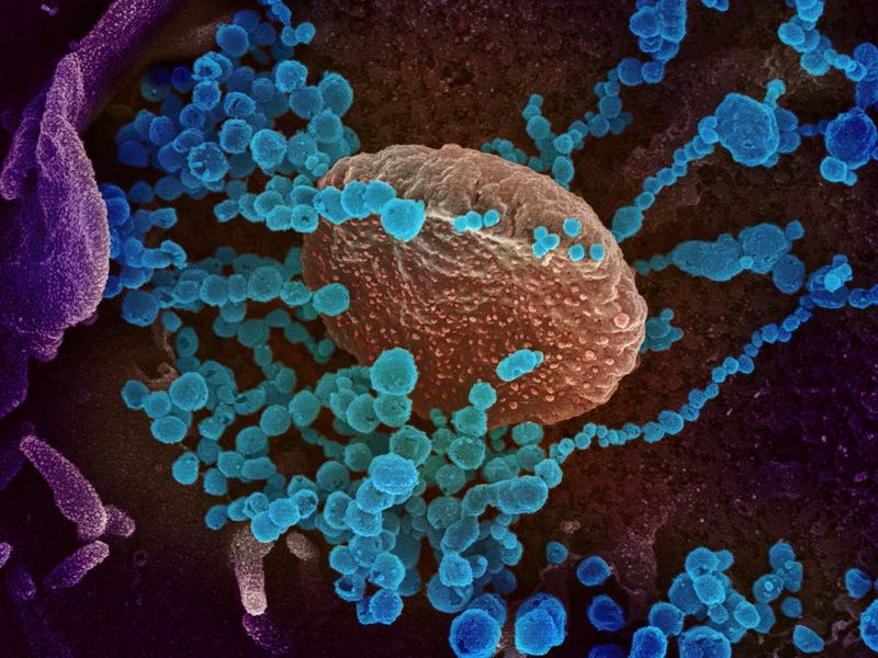
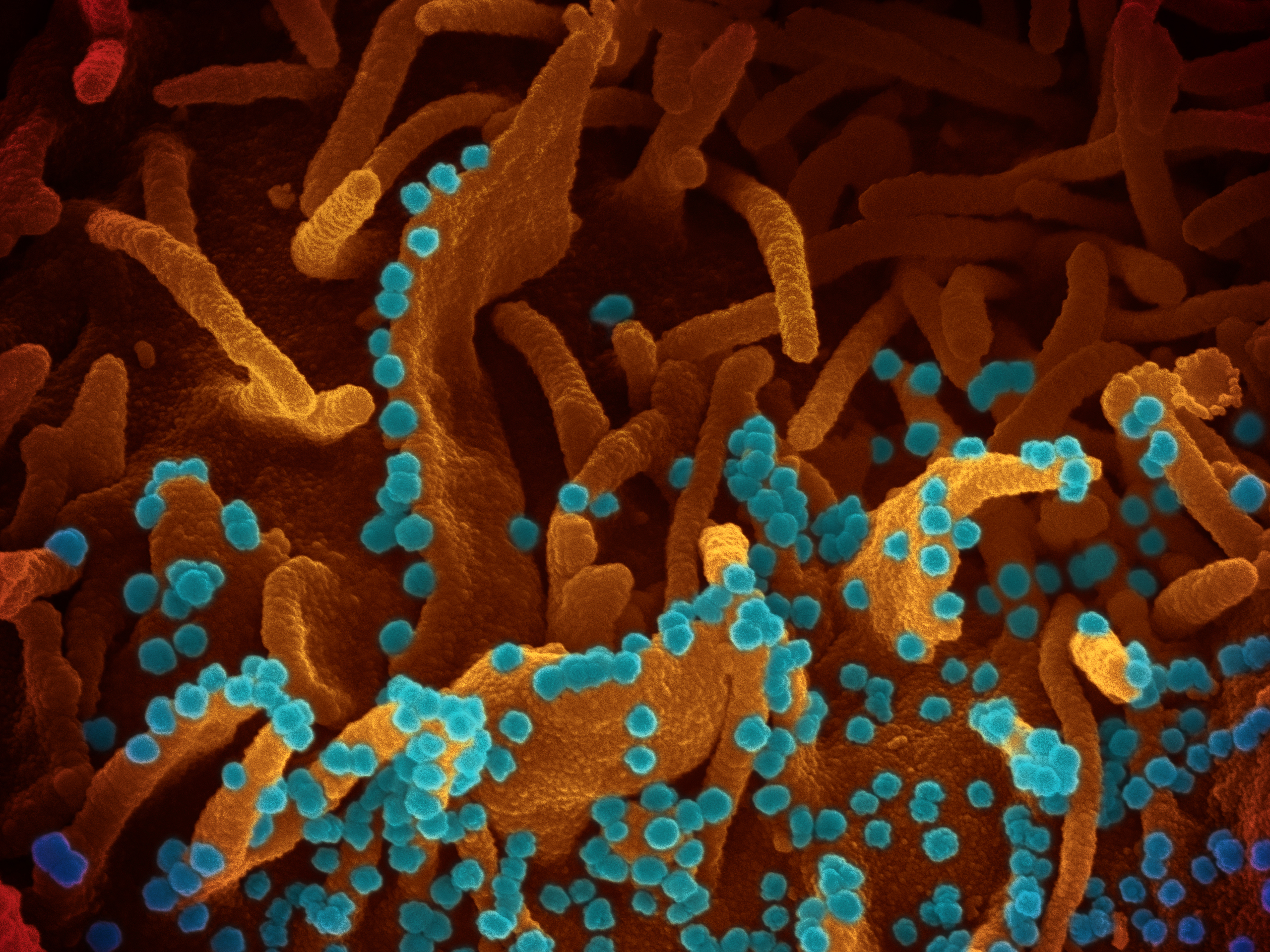
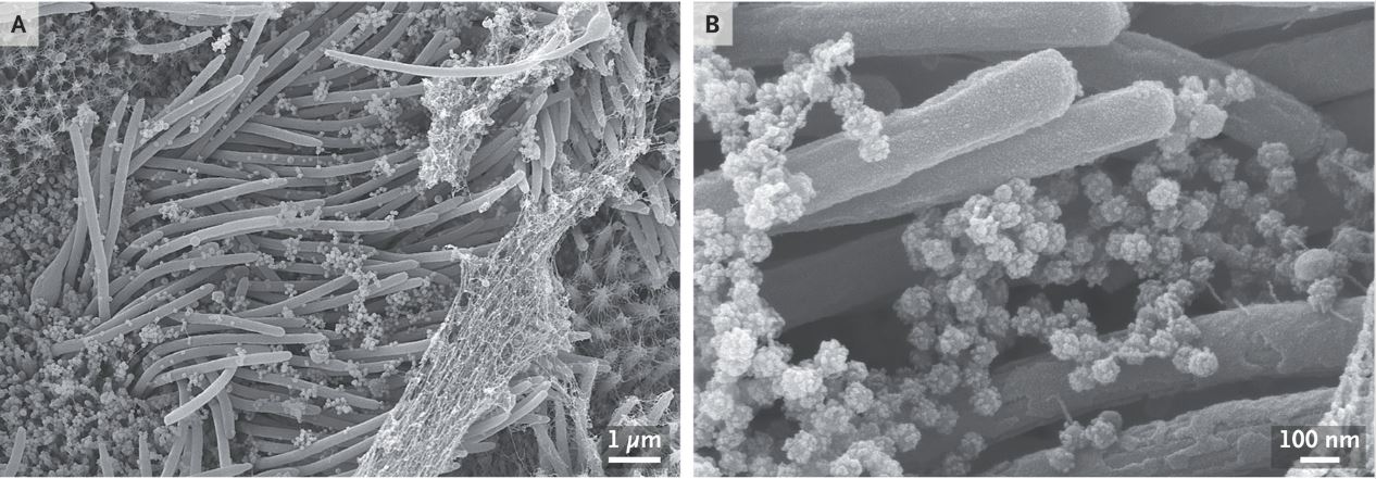 Striking images capture SARS-CoV-2 infecting cells | Live Science
 Microscopy analysis of Zika virus morphogenesis in mammalian cells | Scientific Reports
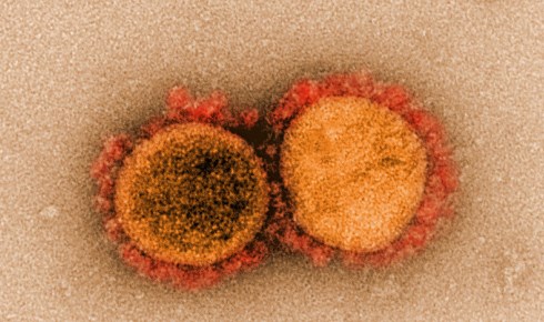
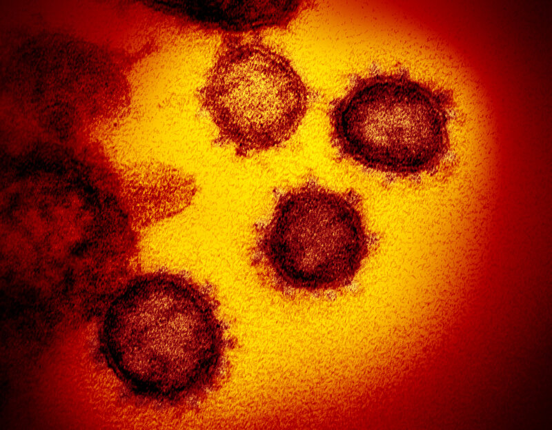 IMAGES: What New Coronavirus Looks Like Under The Microscope : NPR
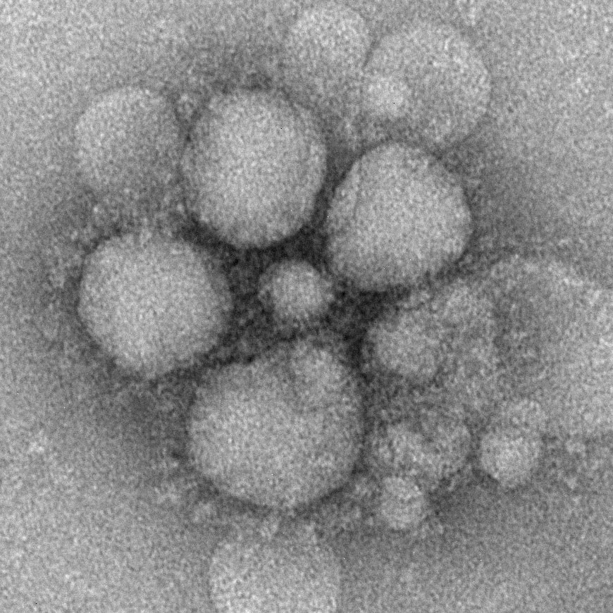
 Electron microscope images of the Zika Virus with a size of around 50... | Download Scientific Diagram
 COVID-19: Portraits of a virus | News-photos – Gulf News
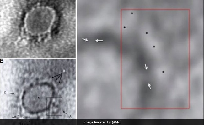 Coronavirus Update: First Electron Microscope Image Of COVID-19 Virus From India Released
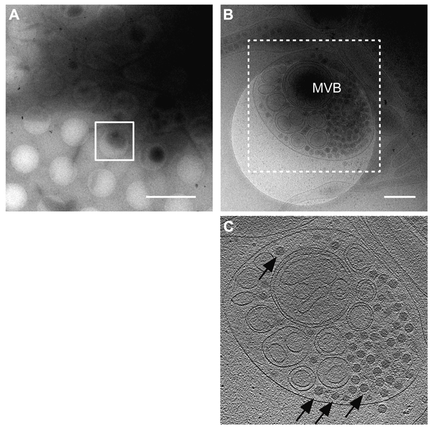 Frontiers | Life cycle of phytoreoviruses visualized by electron microscopy and tomography | Microbiology
 50 Striking Microscopic Images of Viruses and Bacteria | The Weather Channel - Articles from The Weather Channel | weather.com
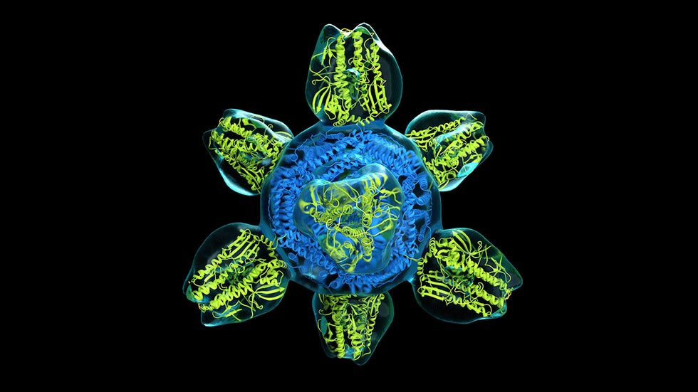
 Cryo-electron microscopy reveals structure of a herpesvirus capsid
 Modern Uses of Electron Microscopy for Detection of Viruses | Clinical Microbiology Reviews
 The Scientist Behind Some of the Best Coronavirus Images | Time
 Modern Uses of Electron Microscopy for Detection of Viruses | Clinical Microbiology Reviews
 Electron Microscopy Images
 Zika virus: Expert discusses its evolution and the challenges of developing effective treatments | Hub
 Hong Kong: first 'proven' case of COVID reinfection | Arab News


Comments
Post a Comment